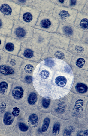
Crossing over can occur over the synaptonemal complex once it has formed, but in some organisms this complex is not obligatory for recombination. Only once the pair has been connected can it be called a tetrad or bivalent. These filaments make up the synaptonemal complex. Ladder-like filaments bring together and attach the chromosome pair at a central point.

In order to attach as a pair, a synapsis is formed. Prophase I Stages Stage 2: ZygoteneĪ tetrad, or two homologous chromosomes consisting of four chromatids, is connected to produce a chromosome pair during meiosis. You will notice the string of beads effect. The image below shows all five stages of Prophase I, starting with leptotene at the top. Leptotene is often named in unison with the following stage as the Leptotene-Zygotene transition, as the first stage is in itself a very short process. Recombination is the result of ‘ crossing over’. Recombination is the result of a process in which the DNA of one chromatid is broken apart and mixed with another non-sister chromatid in order to produce a greater variety of alleles in the offspring. It is also understood that at the leptotene stage, double strand breaks in the DNA occur, preparing for recombination. This gives the string of beads effect, with the unwound DNA giving the appearance of the string, and the wound nucleosomes the beads.Įach chromatid is extremely close to the other and this often gives the effect of a single chromosome. When DNA has been twice wrapped around the core histone, it forms a structure known as the nucleosome. Core histones are the equivalent of sewing-thread spools around which the strand of DNA is coiled. It is therefore packaged using special proteins. If fully stretched out, some DNA may be nearly a centimeter long – much too large for a cell nucleolus. Stage 1: LeptoteneĪt this first stage of Prophase I of meiosis I chromosomes are visible under electron microscopy and look like ‘a string of beads’, where the beads are referred to as nucleosomes. As already mentioned, meiosis I has five separate stages. With a better understanding of the terminology, the complicated process of meiosis is much easier to understand. The X-shape of chromosomes can therefore only be seen at particular stages of cell division. In the absence of cell division, DNA is packed into the nucleus and held together by binding proteins in a much less organized way as chromatin fibers. It is also important to mention that chromosomes are a temporary formation. This allows genetic material to merge upon the fertilization of an egg with sperm, creating a cell containing both parents’ DNA in a diploid cell. Haploid cells only contain half of the genetic information of the parent cell, or ‘n’. Haploid refers to a gamete or sex cell – the spermatozoa in males and ova in females. In the image below, number 1 depicts a single chromatid, 2 shows the centromere that joins both chromatids, 3 is the short (or ‘p’) arm and 4 the long (‘q’) arm of the chromosome.

Each chromosome is made up of two chromatids joined at the middle by a centromere.


 0 kommentar(er)
